Malherbe Calcific Epithelioma: What Is It?
Malherbe calcific epithelioma is also called benign Malherbe epithelioma or pilomatrixoma. It is a benign tumor that occurs mainly in children and young people and is more common in women.
The first references to lesions with calcification are found in the writings of Galen and date back to 200 AD, but the calcific epithelioma was described by Malherbe and Chenantains in 1880. To be exact, the researchers spoke of a calcific epithelioma of sebaceous cells.
In 1949, Lever and Griesemer noted that this tumor often originated in the cells of the hair follicles. In 1965, Cantwell and Reed indicated that it was associated with myotonic dystrophy.
What is Malherbe’s calcific epithelioma?
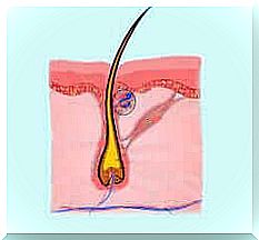
Malherbe’s calcific epithelioma is a neoplasm of an ectodermal nature. It begins in the outermost cells found in the hair follicle sheath. Only very rarely does it become a malignant tumor.
This neoplasm is associated with one or more mutations in the beta-catenin gene (CTNNB1). In 94% of cases it affects the face, neck and upper extremities. In 21% of cases it affects the preorbital region.
More precisely, it has been established that Malherbe’s calcific epithelioma has the following distribution: 30% in the neck, 17% in the cheeks, 16% in the scalp and 14% in the eyebrows. It rarely affects the upper eyelids or lips.
Features
Malherbe’s calcific epithelioma is a tumor that forms in well-defined areas. Usually the dimensions vary from 0.5 to 5 cm in diameter. However, cases with a size of 15 cm have been recorded. The bumps have various shapes and are usually the same color as the skin, or are pink or purplish red.
According to Carbajal and his collaborators, these tumors are present in three classic forms: ulcer-tumor, angioid and ulcer-cutaneous. About 40% of cases occur before the age of 10 and the remaining 60% before the age of 20. A minimum of cases were recorded between the ages of 60 and 70.
These neoplasms most often occur in women. Generally they do not cause pain, but sometimes the person can feel some discomfort. The malignant form is very rare and mainly affects men around the age of 50. These are aggressive tumors with frequent lung metastases.
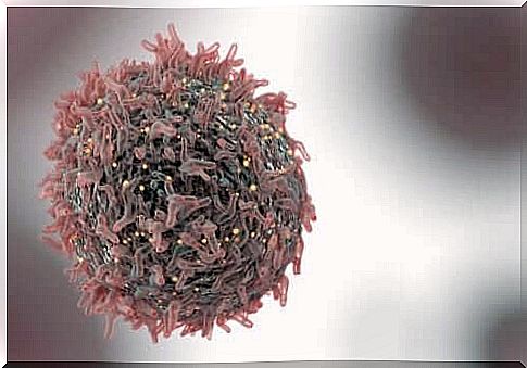
Malherbe’s calcific epithelioma affects two types of cells: basophilic epithelial cells, which account for 50-75% of the tumor; and eosinophilic cells, also called shadow cells or ghost cells, which account for the remaining percentage.
Diagnosis

The diagnosis of Malherbe’s calcific epithelioma is usually clinical. However, a histopathological study is also necessary. Skin biopsy is the method by which a definitive diagnosis is obtained.
Since these neoplasms present a great clinical variety, a differential diagnosis will have to be made. In fact, it is easy to confuse them with other disorders such as dermoid cysts, epidermal cysts and cutaneous calcinosis. They could also be confused with malignant diseases such as giant cell fibroblastoma and others.
Usually, a needle aspiration biopsy (FNA) is done to validate the initial diagnosis. It is recommended that 2 or 3 clinical studies be performed to confirm any previous diagnoses.
Learn more about Malherbe’s calcific epithelioma
The lesion caused by Malherbe’s calcific epithelioma appears as an isolated lump. It has a solid texture and a smooth surface. It moves easily with simple palpation.
To the touch it looks like a pebble with various angled facets. While stretching the skin with your fingers, a hard lump is felt underneath.
In addition to the clinical variants proposed by Carbajal and his collaborators, there are other forms of this tumor such as: lymphangiectasia, exophysiology, perforation and peduncle. A very rare and curious variant is the giant pilomatrixoma which often occurs in subjects suffering from Gardner’s syndrome.